Quick answer
The sesamoid bones are two small bones embedded within the tendon under the big toe joint — the tibial sesamoid (medial) and the fibular sesamoid (lateral). They act like pulleys, redirecting force during push-off. A fracture in either sesamoid is uncommon but real, and is often misdiagnosed as sesamoiditis — the bones look similar enough on X-ray that distinguishing requires careful evaluation. Fractures take longer to heal because the sesamoids have poor blood supply.
The other thing a sesamoid fracture is confused with is a bipartite sesamoid — a normal congenital variant where the sesamoid is naturally in two pieces, present in roughly 10–30% of people. The difference matters: a bipartite sesamoid is harmless and needs no treatment, while a true fracture needs offloading and time to heal. The two can look nearly identical on a single X-ray, and the distinction is the most common diagnostic question with sesamoid two-piece appearance — covered in detail below.
Two types of fracture
Acute fracture
A sudden, traumatic break — typically from:
- Landing forcefully on the ball of the foot from a jump or fall
- High-impact running on a hard surface
- A direct blow to the bottom of the big toe joint
- Forced extension of the big toe (similar mechanism to severe turf toe)
Stress fracture
A gradual fatigue fracture from repetitive load:
- High-mileage runners
- Dancers (especially ballet, en pointe)
- Athletes with rapid increases in training volume
- People with high arches (cavus foot) — concentrates load on the sesamoids
How to distinguish from sesamoiditis
| Feature | Sesamoiditis | Sesamoid fracture |
|---|---|---|
| Onset | Gradual, dull ache | Sudden sharp pain (acute) or persistent sharp ache (stress) |
| Pain quality | Aching | Sharp, focal |
| Tenderness | Diffuse over sesamoid | Pinpoint over the bone |
| Healing time | Weeks to months | Months; sometimes 6–12 months for stress fractures |
| X-ray | Normal | May show fracture line; bipartite sesamoid can mimic |
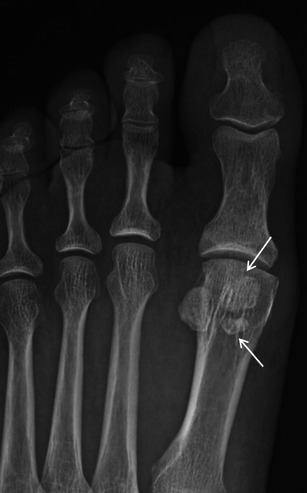
Bipartite sesamoid — the most common mimic
A bipartite sesamoid is a normal anatomic variant in which the sesamoid bone forms in two (or sometimes three — tripartite) pieces rather than one. It is present in roughly 10–25% of people depending on the series, occurs more often in the tibial (medial) sesamoid than the fibular, and is bilateral in about 25–85% of cases. On a standard X-ray, a bipartite sesamoid can look strikingly like a fracture — so much so that telling them apart is one of the more common diagnostic pitfalls in foot radiology.
Features that suggest bipartite (normal variant) rather than fracture:
- Smooth, rounded, well-corticated edges at the split — fractures show sharp, irregular, non-corticated edges
- Total combined size larger than expected — the bipartite halves together are usually bigger than a normal single sesamoid
- No significant pain or tenderness when pressed directly over the sesamoid (although a bipartite sesamoid can develop its own painful problems)
- No clear history of acute injury or escalating overuse
A practical clinical tip: because bipartite sesamoids are bilateral in a large minority of patients, comparing an X-ray of the other foot can help confirm the diagnosis. If the contralateral foot shows the same bipartite pattern, the appearance on the symptomatic side is more likely to be a normal variant than a fracture. (The reverse is not absolute — a unilateral bipartite is still possible — so a symptomatic patient with X-ray findings still needs careful clinical correlation, and MRI when in doubt.)
When the X-ray is genuinely ambiguous, MRI is the most useful next step. A true acute fracture shows bone marrow edema across the line; a quiet bipartite sesamoid does not. A symptomatic bipartite sesamoid (sometimes called bipartite sesamoiditis, where the cartilaginous junction between the two parts becomes inflamed or disrupted) can show edema too — at which point the management overlaps with sesamoiditis and stress fracture care.
How to recognize it
- Sharp, focal pain under the big toe joint
- Pain with push-off, especially during running or walking on hard surfaces
- Tenderness directly over the affected sesamoid
- Swelling and possible bruising under the joint
- Painful big toe extension (bending the toe up stretches the tendon and sesamoid)
- Pain wearing high heels (loads the sesamoid)
- Limp in acute cases
Diagnosis
- History and exam — pinpoint tenderness over the sesamoid, pain with toe extension
- X-rays — including standard views and a sesamoid axial view which best shows the bones. Compare to the other foot if needed
- MRI — gold standard. Shows fracture, bone marrow edema, and helps distinguish from bipartite sesamoid or sesamoiditis
- Bone scan or CT — sometimes used in stress fracture cases
Treatment
Conservative care (most cases)
The sesamoids’ poor blood supply means healing is slow. Treatment focuses on complete unloading of the sesamoid:
- Walking boot or stiff-soled rocker shoe for 6–8 weeks
- Carbon fiber turf toe plate to limit big toe extension
- Sesamoid pad / J-pad orthotic to offload the affected bone
- Activity modification — no running, jumping, or pivoting until cleared
- Crutches in the early phase if pain is severe
- NSAIDs for inflammation
- Bone stimulator — sometimes used in stress fractures with delayed healing
- Imaging follow-up at 6–8 weeks to confirm healing
Healing time:
- Acute fractures: typically 6–12 weeks
- Stress fractures: 3–6 months
- Some cases progress to avascular necrosis (bone death) requiring different management
Surgery
Considered for fractures that don’t heal with prolonged conservative care, or for displaced fractures with poor alignment:
- Sesamoidectomy — removal of the affected sesamoid. Reliable for pain relief
- Bone graft / fixation — preservation surgery, less common but used in selected cases
- Recovery — 6–8 weeks in a boot; full recovery 3–6 months
Removing both sesamoids is generally avoided — it can cause secondary big toe deformity. Single sesamoidectomy is generally well tolerated.
Bottom line
A sesamoid fracture is slow to heal, easy to confuse with sesamoiditis or a bipartite sesamoid, and benefits from early protected weight-bearing. MRI is the most useful imaging test for confirming the diagnosis. Most cases heal with patient conservative care; persistent pain points to sesamoidectomy. Athletes should expect 3–6 months for full return to sport — trying to push through is the most common cause of progression to avascular necrosis.
Frequently asked questions
What is a sesamoid fracture?
A sesamoid fracture is a break in one of the two small bones (about the size of a corn kernel each) embedded in the tendons beneath the big toe joint. The medial sesamoid (tibial sesamoid) is fractured more often than the lateral (fibular). Fractures happen from a single high-force injury (a fall, jump, or stomp landing forefoot-first) or from repetitive overload (stress fracture pattern). Both kinds cause sharp pain under the ball of the foot at the base of the big toe.
Sesamoid fracture vs sesamoiditis — what's the difference?
Sesamoiditis is inflammation of the sesamoid bones and surrounding soft tissue from overuse — common in runners, dancers, and people who spend long periods on the balls of their feet. A sesamoid fracture is an actual break in the bone. The symptoms overlap significantly. The distinguishing test is an X-ray: a fresh fracture line is visible, while sesamoiditis usually shows a normal bone (or sometimes a bipartite sesamoid, which is a normal anatomical variant, not a fracture).
How long does a sesamoid fracture take to heal?
6 to 12 weeks for most cases — sesamoids have a notoriously slow healing pace because of their small size and limited blood supply. Initial treatment is typically a walking boot or cast for 4–6 weeks, followed by a stiff-soled shoe and offloading insert for several more weeks. Recurrent pain or non-union (failure to heal) happens in a meaningful minority of cases and can require either continued conservative care or surgical sesamoidectomy.
Bipartite sesamoid vs sesamoid fracture — how do they look different?
A bipartite sesamoid is a normal variant in which one sesamoid is naturally divided into two parts — present in roughly 10–25% of people and usually painless. The dividing line is smooth, well-corticated, and rounded on X-ray. A fresh fracture line is irregular, sharp-edged, and not corticated. When the appearance is ambiguous, comparison X-rays of the opposite foot help (bipartite sesamoids are often bilateral), and MRI can definitively distinguish acute fracture from a long-standing bipartite.
Do I need surgery for a sesamoid fracture?
Most sesamoid fractures heal with conservative care — protected weight-bearing in a boot, gradual return to a stiff-soled shoe, offloading insert, and patience. Surgery (partial or complete sesamoidectomy) is considered when pain persists for 6+ months despite proper conservative care, when the fracture clearly hasn't healed on imaging, or when there's significant arthritic change. Removing a sesamoid alters big-toe push-off mechanics, so it's not a first-line option.
Sources
Last updated: May 27, 2026

About the author
Written and reviewed by a Doctor of Podiatric Medicine (DPM) practicing in Arizona for 6+ years. Board-certified by the American Board of Podiatric Medicine (ABPM); graduate of Midwestern University Arizona College of Podiatric Medicine.
Last clinically reviewed: May 27, 2026