Quick answer
The peroneal tendons run down the outside of the ankle and help stabilize the foot during lateral movements. Irritation or wear of these tendons causes pain along the outside of the ankle, often after sports or in people with chronic ankle instability. Most cases respond to rest, supportive care, and addressing the underlying mechanics.
Anatomy in plain terms
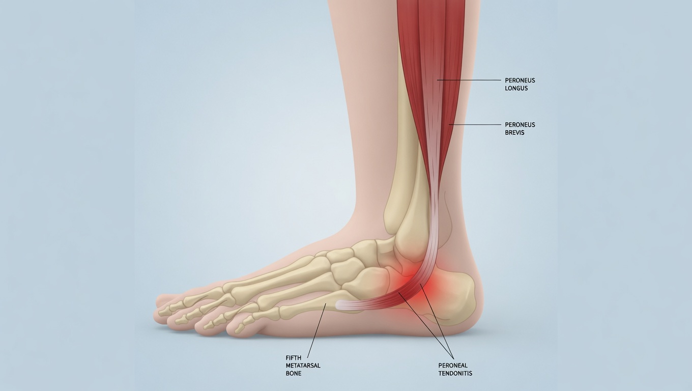
Two tendons share a narrow space behind the lateral malleolus (outside ankle bone):
- Peroneus longus — runs underneath the foot to the inside of the arch
- Peroneus brevis — attaches to the base of the fifth metatarsal (outside of the foot)
Together they:
- Evert the foot (turn the sole outward)
- Stabilize the lateral ankle during walking and especially during lateral cutting
- Protect against sprains by countering inward rolling
When these tendons are overworked or stressed beyond their capacity, they get irritated, develop micro-tears, or partially tear.
How to recognize it
- Pain along the outside of the ankle, behind or below the lateral malleolus
- Pain that radiates down to the side of the foot sometimes
- Worse with activity, particularly running, cutting, jumping
- Worse on uneven surfaces
- Swelling along the outer ankle, sometimes with warmth
- Pain when actively turning the sole outward against resistance
- Tenderness when pressing on the tendons
- Snapping or popping if the tendons are dislocating
Why it happens
Common contributors:
- Repetitive lateral movements (basketball, tennis, soccer, ballet)
- Chronic ankle instability — peroneal tendons work overtime to compensate for loose ligaments
- High arches (cavus foot) — peroneal tendons are stretched farther
- Acute ankle sprains — initial injury can damage the tendons
- Tight calves
- Running on banked surfaces
- Worn-out shoes with poor lateral support
- Sudden increase in activity
The spectrum
Peroneal tendon problems exist on a continuum:
- Tenosynovitis — inflammation of the sheath around the tendons (most common, mildest)
- Tendinosis — chronic degeneration of the tendon itself
- Longitudinal split tear — the tendon develops a lengthwise split (especially peroneus brevis)
- Subluxation/dislocation — the tendons slip out of their normal groove behind the malleolus
- Complete rupture — uncommon, usually from significant trauma
A clinician can often distinguish these with exam, ultrasound, and MRI.
The peroneus longus uses the cuboid bone as a pulley as it crosses the bottom of the foot. When that pulley relationship gets disrupted, lateral midfoot pain that mimics peroneal tendinitis can develop — see cuboid syndrome for the differential.
Diagnosis
- Physical exam — tenderness location, resisted eversion strength, swelling
- Ultrasound — visualizes the tendons; can detect tears and dislocation in motion
- MRI — gold standard for soft tissue detail
- X-rays — rule out fractures, especially fifth metatarsal injuries
Treatment
Conservative care (first-line for most)
- Reduce aggravating activity — temporary cessation of sports or lateral movements
- Ice after activity for 15–20 minutes
- NSAIDs short-term for pain
- Activity modification — switch to low-impact training (swimming, cycling)
- Supportive footwear with good lateral stability
- Ankle brace during activity
- Custom orthotics — particularly for high-arched feet, with a lateral wedge
- Physical therapy — eccentric strengthening, calf flexibility, balance work
When more is needed
- Walking boot for 2–4 weeks for severe inflammation or partial tear
- Cortisone injection — used cautiously; can weaken tendons
- Platelet-rich plasma (PRP) injection — newer option, mixed evidence
Surgery
For tears that fail conservative care, or for tendon dislocation:
- Tear repair — direct repair or tubularization
- Groove deepening — for dislocation, deepens the groove behind the malleolus
- Retinacular reconstruction — for chronic dislocation
- Recovery typically 8–12 weeks; full return to sports 4–6 months
Bottom line
Peroneal tendon issues are common in athletes and in people with chronic ankle instability. Most cases respond to rest, eccentric strengthening, supportive shoes, and addressing the underlying mechanical contributors. Persistent or severe symptoms warrant imaging to look for tears or dislocation that may need more focused treatment.
Last updated: April 25, 2026

About the author
Written and reviewed by a Doctor of Podiatric Medicine (DPM) practicing in Arizona for 6+ years. Board-certified by the American Board of Podiatric Medicine (ABPM); graduate of Midwestern University Arizona College of Podiatric Medicine.
Last clinically reviewed: April 25, 2026