Quick answer
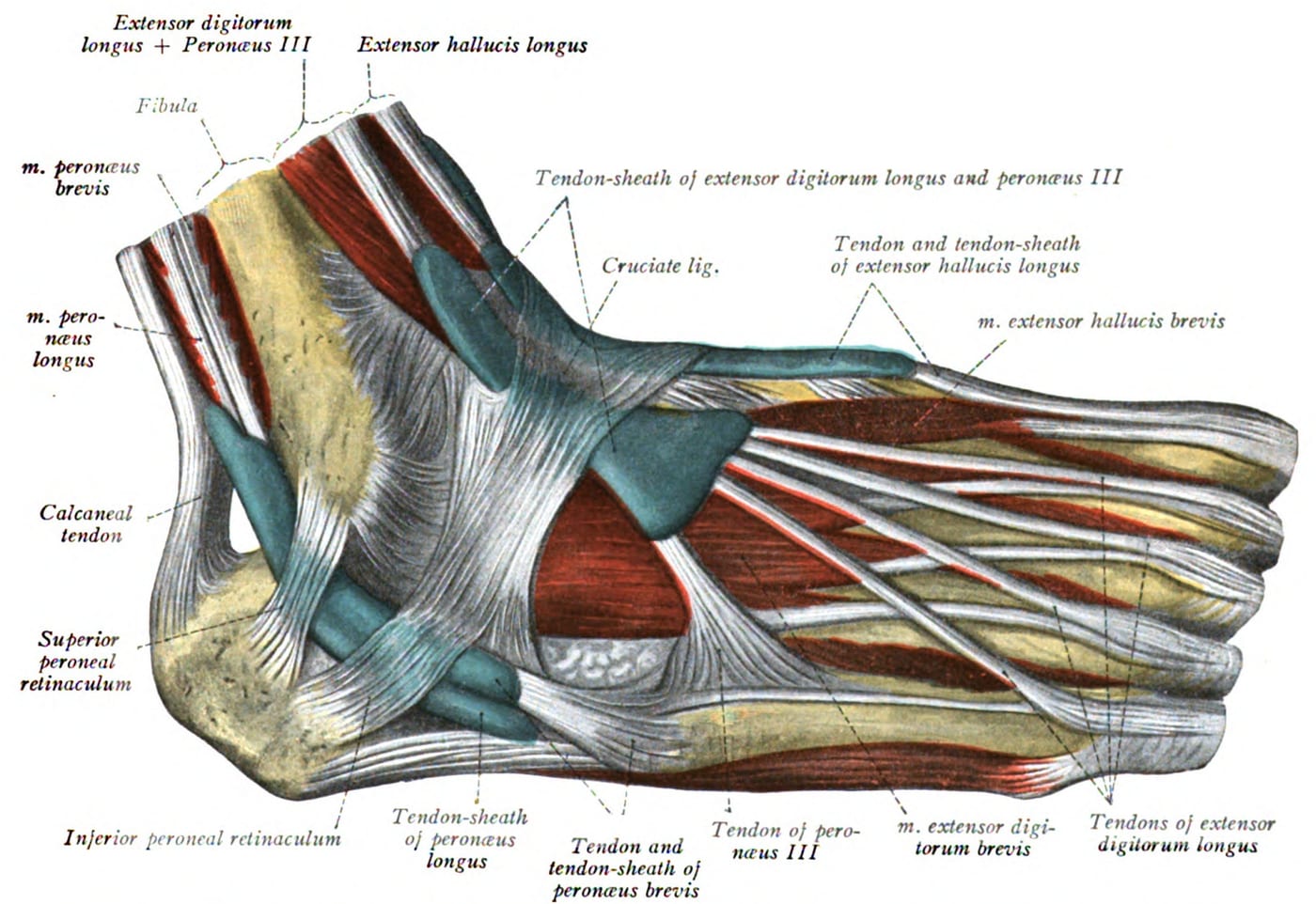
The two peroneal tendons — peroneus longus and peroneus brevis — run together behind and below the lateral ankle bone (lateral malleolus). They evert the foot and stabilize the ankle. A tear in one of these tendons is a common but frequently overlooked source of chronic lateral ankle pain. It’s not the same thing as peroneal tendinitis (irritation without tearing) — a tear is structural damage that often needs more than rest.
How tears happen
- Acute tear — usually with a high-energy ankle sprain. The peroneus brevis is most often affected, often as a longitudinal split rather than a clean transverse tear
- Chronic / attritional tear — gradual fraying of the tendon as it rubs against the back edge of the fibula or against a bony prominence. Common contributors:
- Chronic ankle instability — repeated rolling stresses the tendon
- High arch (cavus) foot — turns the heel inward and increases peroneal tendon load
- Os peroneum — an extra small bone in the peroneus longus that can fracture or cause attritional tearing
- Subluxation — the tendons slipping out of their groove (a related but separate problem)
The peroneus brevis tears are often longitudinal splits (the tendon splays into two slips), while peroneus longus tears tend to be more discrete tears, sometimes associated with os peroneum problems.
How to recognize it
- Persistent pain along the outer ankle behind and below the lateral malleolus
- Swelling along the tendon path
- Pain with eversion against resistance
- Snapping or popping with ankle motion (more typical of subluxation but possible with tear)
- Weakness when pushing off to the outside
- Often a history of repeated sprains or chronic ankle instability
- Pain that doesn’t fit a simple ligament sprain and persists weeks to months
Diagnosis
- Physical exam — tender along the tendons, pain with resisted eversion, sometimes a palpable thickening
- Ultrasound — dynamic and inexpensive; shows tears and subluxation
- MRI — gold standard for tendon detail, identifying tear length, location, and associated problems
- Weight-bearing X-rays — to assess foot alignment (especially for high arch); identify os peroneum or fractures
Treatment
Conservative care
- Rest, NSAIDs, and walking boot for 4–6 weeks during the acute phase
- Lateral wedge orthotics to offload the peroneal tendons in cavus (high arch) feet
- Physical therapy for peroneal strengthening and balance
- Bracing for chronic instability cases
- Cortisone injection — generally avoided around tendons because of rupture risk
Conservative care helps for partial tears, especially when underlying mechanics (high arch, instability) are addressed. It’s less effective for complete tears or large longitudinal splits.
Surgery
For tears that fail conservative care, or for athletes who need to return to sport:
- Tubularization / repair — sewing a longitudinal split back together
- Debridement of frayed edges
- Tendon transfer — if one tendon is unsalvageable, the other peroneal can be transferred
- Cavus correction — if a high arch is the underlying driver, a calcaneal osteotomy may be added to redirect forces
- Recovery — typically 2–6 weeks non-weight-bearing or in a boot, then progressive rehab over 3–6 months
Bottom line
A peroneal tendon tear is one of the most commonly missed causes of chronic outer ankle pain. Anyone with persistent symptoms after a sprain — especially with multiple sprains, instability, or high arches — should be evaluated specifically for peroneal pathology. MRI is the most useful study. Underlying mechanics (instability, high arch) need to be addressed alongside the tendon itself, or recurrence is common.
Last updated: April 27, 2026

About the author
Written and reviewed by a Doctor of Podiatric Medicine (DPM) practicing in Arizona for 6+ years. Board-certified by the American Board of Podiatric Medicine (ABPM); graduate of Midwestern University Arizona College of Podiatric Medicine.
Last clinically reviewed: April 27, 2026