
Quick answer
The Achilles tendon connects the calf muscles to the heel bone and is the largest tendon in the body. A rupture means it tears completely. The classic story: a middle-aged athlete pushes off hard during basketball, tennis, or pickleball — feels a sudden “pop” or as if someone kicked the back of their leg — looks behind them, sees no one there. They can often still walk, but pushing off feels weak. About 25% of ruptures are missed on initial evaluation because surrounding muscles partially compensate.
Why it happens
Most ruptures occur in a tendon that already has microscopic damage from years of use:
- The blood supply to the Achilles is poorest about 2–6 cm above the heel — the most common rupture site
- Sudden eccentric load (push-off, jumping, sprinting) overwhelms the weakened area
- Risk factors: prior Achilles tendinopathy, fluoroquinolone antibiotics (ciprofloxacin, levofloxacin), corticosteroid injections near the tendon, sudden increase in activity, advanced age
Less commonly, ruptures result from direct trauma (laceration) or, rarely, in young athletes with no prior issues.
How to recognize it
- Sudden “pop” or kick sensation in the back of the calf
- Weakness with push-off — going up on the toes, climbing stairs
- A palpable gap in the tendon about 2–6 cm above the heel
- Bruising and swelling that develops over hours
- Positive Thompson test — when the calf is squeezed, the foot doesn’t move (in a healthy tendon, the foot points downward)
- Walking is still possible because other muscles (tibialis posterior, peroneals, FHL) can produce some plantarflexion. This is why ruptures get missed.
Diagnosis
- Clinical exam — Thompson test plus a palpable gap is highly accurate
- Ultrasound — quick, inexpensive, and confirms the tear and gap size
- MRI — when chronic tears are suspected or surgical planning needs more detail
Treatment
Treatment choice depends on the patient’s age, activity level, and the size and location of the tear.
Non-surgical (functional rehabilitation)
Modern non-surgical protocols use a walking boot with progressive heel wedges and early functional rehabilitation. Outcomes are nearly equivalent to surgery for many patients, with lower complication rates:
- Initial immobilization in plantarflexion (toes pointed down) to bring tendon ends together
- Progressive dorsiflexion over 6–8 weeks
- Weight-bearing as tolerated in the boot starting around 2 weeks
- Physical therapy focused on calf strength and gait
- Return to sport typically 6–9 months
- Re-rupture rate is slightly higher than with surgery in some studies, but recent protocols have narrowed the gap
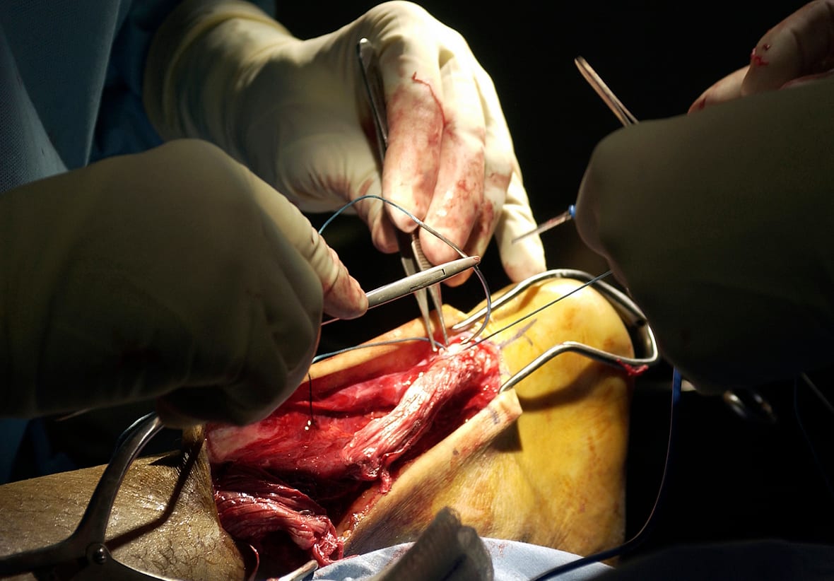
Surgical repair
- Open or percutaneous repair — the tendon ends are sewn back together
- Faster return to sport (4–6 months vs 6–9)
- Lower re-rupture rate historically, though gap is narrowing
- Higher complication rate — wound healing problems, sural nerve injury, infection
- Generally favored for elite athletes and large gaps
Recovery — both paths
- Months 1–2: walking boot, progressive weight-bearing
- Months 2–4: out of boot, range of motion and strength work
- Months 4–6: progressive return to running and sport-specific drills
- Months 6+: return to cutting and jumping sports
When to seek care — and how urgently
Same-day evaluation (emergency department, urgent care, or same-day orthopedic / podiatric clinic) for any of these:
- A sudden “pop” in the back of the calf or heel during sport, push-off, or sudden movement
- The sensation that someone kicked or hit you in the back of the leg, when no one was there
- New weakness with push-off — difficulty going up on the toes or climbing stairs
- A palpable gap in the back of the calf 2–6 cm above the heel
- Bruising or swelling behind the heel after a sudden injury
Do not “walk it off” or wait until next week. Walking is often still possible after an Achilles rupture because other muscles partially compensate — that is precisely how about a quarter of ruptures get missed at first encounter. Outcomes are best when treatment begins within the first 1–2 weeks (operative or non-operative), because the tendon ends are still close together and have not retracted or scarred apart. Late-presenting ruptures are harder to repair, more often require tendon transfers or grafts, and have worse functional outcomes.
Bottom line
An Achilles rupture is a dramatic but treatable injury — if it is recognized promptly. The most important step is early, in-person diagnosis — same-day evaluation if a “pop” was felt, even if you can still walk. Treatment paths (operative vs non-operative) have similar long-term outcomes for most patients, so the decision is individualized between you and your foot-and-ankle surgeon. Either way, expect a 6–9 month return to full sport. Returning too early is the most common cause of re-rupture. This page is general information; the diagnosis and treatment plan need to come from a clinician examining your leg in person.
Last updated: April 27, 2026

About the author
Written and reviewed by a Doctor of Podiatric Medicine (DPM) practicing in Arizona for 6+ years. Board-certified by the American Board of Podiatric Medicine (ABPM); graduate of Midwestern University Arizona College of Podiatric Medicine.
Last clinically reviewed: April 27, 2026