Quick answer
The navicular is a small but structurally critical bone at the apex of the medial arch, sitting between the talus (ankle bone) and the three cuneiform bones of the midfoot. Acute traumatic navicular fractures occur with direct crushing force, high-energy twisting, or ligament avulsion during an ankle sprain. They are less common than navicular stress fractures and are frequently missed when the injury is attributed to a bad sprain.
Note: This page covers acute (traumatic) navicular fractures. The more common navicular stress fracture — a gradual-onset injury in athletes from repetitive loading — is covered on a separate page.
Fracture types
Avulsion fractures (most common) A small chip of bone is pulled away by the tibionavicular ligament (part of the deltoid) or talonavicular ligament during a forceful ankle roll. These are the mildest type — essentially identical in mechanism to an ankle sprain, but with a bony avulsion at the navicular rather than pure ligament tearing.
Navicular body fractures A direct break through the body of the bone, usually from compression or a crushing force to the midfoot. These are more serious and may involve the talonavicular or naviculocuneiform joints.
Navicular tuberosity fractures A specific avulsion fracture where the posterior tibial tendon pulls off a fragment at the navicular tuberosity (the bony prominence on the inner ankle). Common in adults with flatfoot deformity who sustain a twisting injury; also seen in patients with an accessory navicular bone.
How to recognize one
Classic history: A twisting fall or midfoot crush. Avulsion types can look identical to an ankle sprain — the key differentiator is where the pain is. Navicular pain is on the top-inner midfoot, not over the lateral ankle bones.
Symptoms:
- Tenderness over the navicular (the bony prominence on the inner midfoot, just in front of the ankle)
- Swelling and bruising across the midfoot and medial ankle
- Pain with weight-bearing
- Pain with toe push-off or arch-loading activities
How it is diagnosed
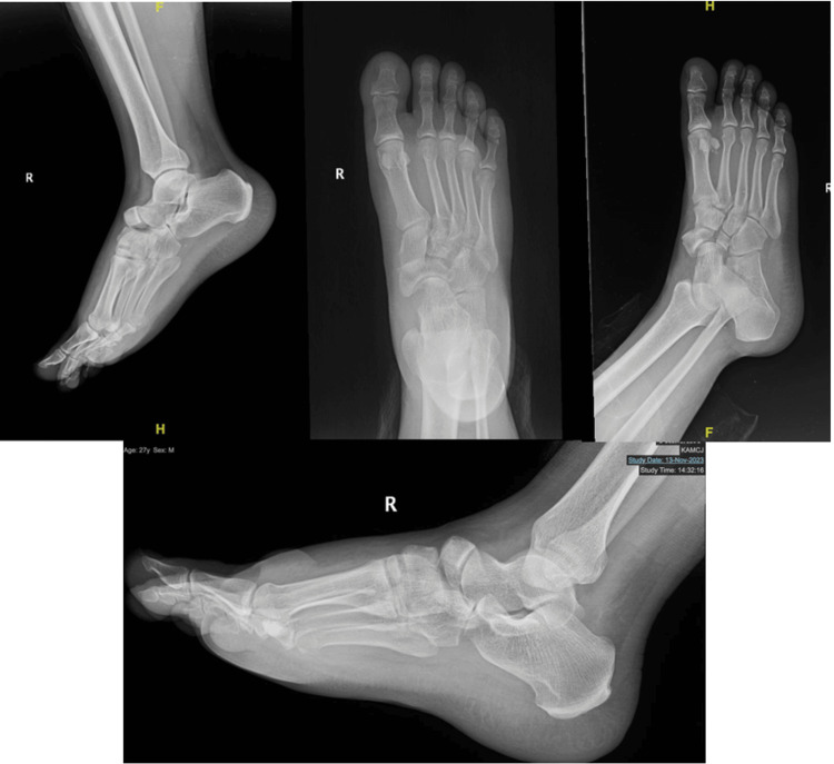
X-ray: Standard three-view foot X-rays will show most navicular fractures. Avulsion chips are small and may be subtle. Body fractures are more apparent.
CT scan: Recommended for all suspected navicular body fractures to assess joint involvement, displacement, and comminution — essential for surgical planning.
MRI: Best for early stress fractures (covered separately) and for evaluating associated soft tissue injuries (posterior tibial tendon, spring ligament, deltoid ligament).
The Lisfranc connection
Navicular body fractures often occur as part of a Lisfranc-type midfoot injury complex. If a navicular fracture is found, the entire tarsometatarsal (Lisfranc) joint complex should be evaluated — including weight-bearing X-rays looking for widening between the 1st and 2nd metatarsal bases or between the medial and middle cuneiform.
A missed Lisfranc injury can leave permanent instability and post-traumatic arthritis.
Treatment
Non-surgical
Avulsion and tuberosity fractures (non-displaced):
- Walking boot or below-knee cast for 4–6 weeks
- Protected weight-bearing as tolerated
- Transition to supportive footwear with arch support
Surgical
Displaced navicular body fractures or fractures involving the talonavicular or naviculocuneiform joint:
- Open reduction and internal fixation (ORIF) with screws to restore joint alignment
- Bone graft may be needed for comminuted fractures
- Goal is to restore arch height and joint congruence
Navicular tuberosity fractures with posterior tibial tendon involvement:
- Surgical repair may be indicated if the tendon’s function is compromised, particularly in patients with preexisting flatfoot
Recovery
| Type | Boot/cast | Return to activity |
|---|---|---|
| Avulsion fracture | 4–6 weeks | 8–10 weeks |
| Tuberosity fracture | 6–8 weeks | 10–12 weeks |
| Body fracture (non-surgical) | 8–10 weeks | 3–4 months |
| Body fracture (surgical) | 10–12 weeks NWB, then boot | 5–8 months |
Post-traumatic arthritis in the talonavicular joint is a recognized late complication, particularly after displaced body fractures or joint involvement.
The main thing to understand
Any midfoot injury with significant pain, swelling, or difficulty bearing weight needs in-person evaluation by a clinician — and X-rays are essential. Acute navicular fractures — particularly avulsion types — are easily mistaken for ankle sprains, and the consequences of missing them can be permanent: chronic pain, post-traumatic arthritis, and unrecognized Lisfranc injury. The distinguishing clinical feature — pain over the navicular rather than the lateral ankle ligaments — is suggestive but not definitive. Imaging (initial three-view foot X-rays, often followed by CT or MRI) is the only reliable way to confirm whether there is a fracture, classify it, and screen for the associated Lisfranc complex injuries that change management.
This is not an injury to manage from a website. Same-day evaluation by a foot and ankle clinician (orthopedic or podiatric surgeon, urgent care, or emergency department) is the right next step if you have midfoot pain after an injury — even if you can still walk on it. Treatment decisions (boot vs. cast vs. surgery, weight-bearing vs. non-weight-bearing) need to come from a clinician who has seen your X-rays and examined your foot in person.
Frequently asked questions
How serious is a navicular fracture?
A navicular fracture is taken seriously because the navicular is the keystone bone of the midfoot — it links the hindfoot to the forefoot and helps maintain the arch. Acute fractures from high-energy injuries (falls, car accidents) often involve other midfoot bones and can lead to long-term arthritis or arch collapse if not treated properly. Acute avulsion fractures (small chip fractures at tendon attachments) are usually less serious and heal with a boot.
Navicular fracture vs navicular stress fracture — how do they differ?
An acute navicular fracture happens from a single high-energy event — a car accident, fall from height, or direct blow. A navicular stress fracture develops gradually from repetitive overload, classically in distance runners and jumping athletes. Stress fractures are notorious for being missed on initial X-rays (the line is subtle or invisible) and often require MRI or CT for diagnosis. Both heal slowly because of the navicular's limited blood supply.
How long does a navicular fracture take to heal?
Healing depends on the fracture type and treatment. Non-displaced acute fractures typically heal in 6 to 8 weeks of non-weight-bearing cast or boot. Displaced or comminuted fractures usually require surgical fixation followed by 8–12 weeks of non-weight-bearing and then progressive weight-bearing. Avulsion (chip) fractures heal faster, often 4–6 weeks in a boot. Navicular stress fractures take 8–12 weeks minimum and have a high non-union rate if treated too casually.
Does a navicular fracture need surgery?
Non-displaced acute fractures are usually treated with cast immobilization and non-weight-bearing. Displaced fractures (more than 1–2 mm of displacement, particularly involving the joint surface) typically need surgical fixation with screws to restore alignment and prevent long-term arthritis. Stress fractures often require surgery if they involve the high-risk central one-third zone of the bone, especially in athletes who want a faster, more reliable return to sport.
Why is the navicular often missed on X-ray?
The navicular sits in the middle of an overlapping cluster of midfoot bones, and stress fracture lines through it can be very subtle — sometimes invisible on initial X-rays. Acute fractures with displacement are usually obvious, but navicular stress fractures are one of the most commonly missed midfoot fractures. Clinicians who suspect navicular involvement should request additional views and have a low threshold for ordering MRI or CT, especially in distance runners or jumping athletes.
Last updated: April 26, 2026

About the author
Written and reviewed by a Doctor of Podiatric Medicine (DPM) practicing in Arizona for 6+ years. Board-certified by the American Board of Podiatric Medicine (ABPM); graduate of Midwestern University Arizona College of Podiatric Medicine.
Last clinically reviewed: April 26, 2026