Quick answer
The midfoot has multiple joints — the joints between the cuneiforms, navicular, cuboid, and metatarsal bases. Together they form a relatively rigid arch that transfers force during walking. Midfoot arthritis affects these joints, most commonly the tarsometatarsal (Lisfranc) joints between the cuneiforms and the metatarsals. Patients typically describe pain across the top of the midfoot that’s worse with walking, especially with push-off, and a visible bump may develop on the top of the foot.
Causes
- Post-traumatic (most common): Following a Lisfranc injury, even when the injury is well-treated. Cartilage damage at the time of injury, plus altered joint mechanics, drive arthritis over years
- Primary osteoarthritis: Wear-and-tear from cumulative loading. More common in older adults, often runs in families
- Inflammatory arthritis: Rheumatoid, psoriatic, gout
- Progressive flatfoot: Long-standing arch collapse stresses the midfoot joints
- Charcot foot: Severe destructive arthropathy in diabetic patients with neuropathy
How to recognize it
- Pain across the top of the midfoot, often described as a deep, aching pain
- Worse with walking, particularly with push-off and on uneven ground
- Better with rest
- Visible “dorsal bump” — bone spurs on the top of the foot (a hallmark of midfoot OA)
- Difficulty fitting in lace-up shoes because of the bump and lace pressure
- Stiffness in the midfoot, especially in the morning
- A sense the foot “doesn’t push off right” during running or athletic activity
Diagnosis
- History and exam — tenderness over specific joints; pain reproduced with passive twisting of the midfoot
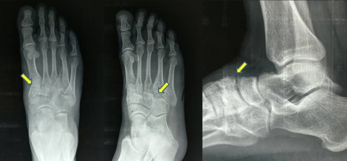
- Weight-bearing X-rays — show joint space narrowing, dorsal osteophytes, and any old Lisfranc injury patterns. Multiple views (AP, lateral, oblique) help identify which joints are involved
- CT scan — gold standard for assessing joint surfaces, especially for surgical planning
- MRI — useful for early disease and soft tissue assessment
- Diagnostic injections — placing local anesthetic into specific joints can confirm which joint is generating pain (multiple midfoot joints often look arthritic on imaging, but only one or two are symptomatic)
The last point matters: only the symptomatic joints need surgical attention, even if X-rays show widespread changes.
Treatment
Conservative care (first-line)
Most patients can be managed non-operatively for years:
- Stiff-soled shoes — limit midfoot motion and reduce pain. Hiking boots or shoes with carbon-fiber inserts work well
- Custom orthotics — with a midfoot post to support and offload arthritic joints
- NSAIDs for inflammation flares
- Lacing modifications to reduce pressure over the dorsal bump — skip eyelets over the prominence
- Activity modification — substitute lower-impact exercise during flares
- Cortisone injections — into specific arthritic joints. Often very effective; can be repeated periodically
- Weight loss when relevant
Surgery
For patients who fail conservative care:
- Midfoot fusion (arthrodesis) — the standard. Specific joints (rarely all) are fused. Pain relief is typically excellent because the midfoot is normally a relatively rigid structure with minimal motion to lose
- Dorsal exostectomy — removal of the dorsal bone spur alone. Used in selected cases when the bump is the main problem
- Recovery — typically 6–8 weeks non-weight-bearing in a cast or boot, then progressive return to weight-bearing over 3–6 months. Full recovery 6–12 months
- Outcomes — patient satisfaction with midfoot fusion is generally high when properly indicated
The trade-off: very modest loss of midfoot motion (which the average patient doesn’t notice) for significant pain relief.
Bottom line
Midfoot arthritis is the classic delayed consequence of a Lisfranc injury but also occurs as primary OA. Most patients do well with stiff shoes, orthotics, and selective injections for years. Midfoot fusion is reliable when surgery is needed because the joints involved have little normal motion to begin with. Diagnostic injections are key to picking which joints to target — both for confirming the source of pain and for surgical planning.
Last updated: April 27, 2026

About the author
Written and reviewed by a Doctor of Podiatric Medicine (DPM) practicing in Arizona for 6+ years. Board-certified by the American Board of Podiatric Medicine (ABPM); graduate of Midwestern University Arizona College of Podiatric Medicine.
Last clinically reviewed: April 27, 2026