Quick answer
A Lisfranc injury affects the tarsometatarsal joints in the middle of the foot — where the metatarsals meet the midfoot bones. It spans a wide spectrum from a mild ligament sprain to a severe fracture-dislocation. It’s notoriously easy to miss because patients can sometimes still walk after the injury — but missing it can lead to permanent midfoot arthritis.
What’s actually injured
The midfoot is held together by a complex of ligaments connecting the cuneiforms (small midfoot bones) to the metatarsals. The most important is the Lisfranc ligament, which runs from the medial cuneiform to the base of the second metatarsal — anchoring the entire midfoot arch.
When this ligament tears (and often associated bones fracture), the midfoot loses its stability. The metatarsals can shift relative to the cuneiforms, and the arch can collapse — sometimes immediately, sometimes over months as the foot adapts to instability.
How to recognize it
The classic story: a foot that’s planted with the toes pointed downward (a “tip-toe” position) when a force comes down on the back of the heel, or a midfoot twist during sports. After the injury, the patient can sometimes still walk — which is why this is so often dismissed as a “bad sprain.”
Key features:
- Midfoot pain and swelling out of proportion to a “minor” injury
- Plantar ecchymosis — bruising on the bottom of the midfoot. This is the single most specific sign of a Lisfranc injury.
- Pain with passive twisting of the midfoot
- Inability to bear weight — or pain so significant that walking is markedly limited
- Midfoot widening in severe cases
- Persistent pain after a “minor” twist that doesn’t improve in 1–2 weeks
The spectrum of injury
Lisfranc injuries range from subtle to dramatic:
- Sprain (stable) — partial ligament tear, joints still aligned. Heals with immobilization.
- Sprain (unstable) — complete ligament tear, joints subtly malaligned with weight bearing. Often missed without weight-bearing X-rays.
- Fracture-dislocation — broken bones plus dislocation. Obvious on X-ray, severe trauma.
The middle category — unstable sprain without obvious fracture — is the most commonly missed and the most important to catch.
Why missed Lisfranc injuries are a problem
When unstable Lisfranc injuries aren’t surgically stabilized:
- The midfoot collapses progressively over months to years
- The arch flattens
- Post-traumatic arthritis develops in the tarsometatarsal joints
- Chronic midfoot pain with walking
- Eventual fusion surgery may be needed to relieve symptoms
Catching the injury early and stabilizing it prevents this cascade.
Diagnosis
This is one of the most important — and most often delayed — diagnoses in foot trauma:
- Physical exam — midfoot tenderness, plantar bruising, pain with passive forefoot abduction
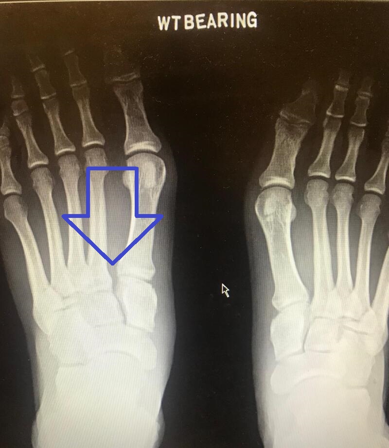
- Weight-bearing X-rays — essential. Non-weight-bearing X-rays can miss subtle Lisfranc injuries entirely. The clue: a gap between the first and second metatarsal bases (>2 mm), or a small “fleck sign” representing an avulsion fragment.
- Comparison views of the uninjured foot can highlight subtle differences
- CT scan — for fracture detail
- MRI — for ligament injury when X-rays are normal but suspicion remains
If a patient has midfoot bruising on the bottom of the foot and continues to have pain after a twist, weight-bearing X-rays should always be obtained.
Treatment
Stable (mild) sprains
- Walking boot or cast for 4–6 weeks, non-weight-bearing initially
- Repeat weight-bearing X-rays at 2 weeks to ensure stability is maintained
- Gradual return to weight bearing in supportive footwear
- Physical therapy for strength and proprioception
- Return to sport typically 3–4 months
Unstable injuries (most cases)
Surgery is generally recommended:
- Open reduction with internal fixation (ORIF) — screws and/or plates anatomically reduce the joints. Hardware is sometimes removed later.
- Primary arthrodesis (fusion) — permanently fuses the affected tarsometatarsal joints. Increasingly favored for severe ligamentous injuries; eliminates the risk of post-traumatic arthritis.
- Recovery typically 6–12 months for return to sport
- Outcome depends heavily on early, accurate diagnosis — late surgery has poorer results
The choice between ORIF and primary fusion depends on whether the injury is primarily bony (favors ORIF) or ligamentous (favors fusion).
Bottom line
Lisfranc injuries are one of the most commonly missed foot injuries, and missing them is costly — chronic pain and arthritis are common consequences. Any midfoot pain after a twist injury — especially with bruising on the bottom of the foot or persistent weight-bearing pain — deserves weight-bearing X-rays and careful evaluation. Early, accurate diagnosis is the single biggest factor in good long-term outcomes.
Last updated: April 25, 2026

About the author
Written and reviewed by a Doctor of Podiatric Medicine (DPM) practicing in Arizona for 6+ years. Board-certified by the American Board of Podiatric Medicine (ABPM); graduate of Midwestern University Arizona College of Podiatric Medicine.
Last clinically reviewed: April 25, 2026