
Quick answer
Charcot foot is a progressive collapse of the bones of the foot, almost always in someone with diabetes who’s lost sensation in their feet from diabetic neuropathy. It starts as a red, hot, swollen foot, often mistaken for an infection or sprain. Without prompt treatment, the foot deforms permanently into a “rocker bottom” shape that’s prone to ulcers and amputation. Catching it early matters enormously.
Why this is so urgent
Two things make Charcot foot a true emergency:
- It’s frequently missed: the red hot swollen foot mimics cellulitis, sprain, or DVT
- The window for preventing permanent deformity is narrow: typically a few weeks
The combination of “easy to miss” plus “narrow treatment window” is why awareness matters.
How to recognize it
The early (active) stage:
- Red, hot, swollen foot in someone with diabetic neuropathy
- Often without significant pain: because the nerves are damaged
- Usually one foot only
- Skin is warm to the touch: typically 2–3°C warmer than the other foot
- Often no clear injury the patient can recall
- Pulses are intact (unlike with arterial disease)
- No fever or systemic illness typically (unlike infection)
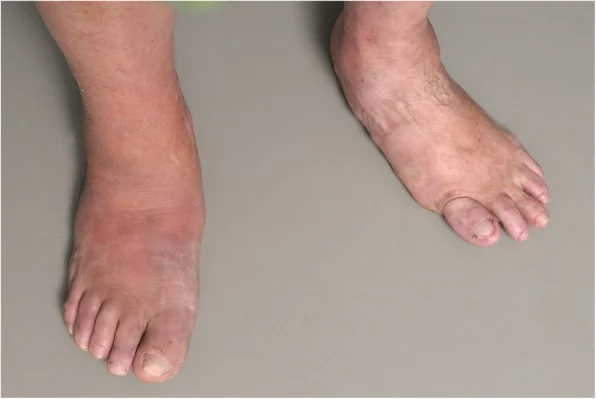
The chronic (later) stage:
- Foot has collapsed into a flat or “rocker bottom” shape
- Deformity makes shoes fit poorly
- High-pressure points develop ulcers under bony prominences
- Risk of infection and amputation climbs significantly
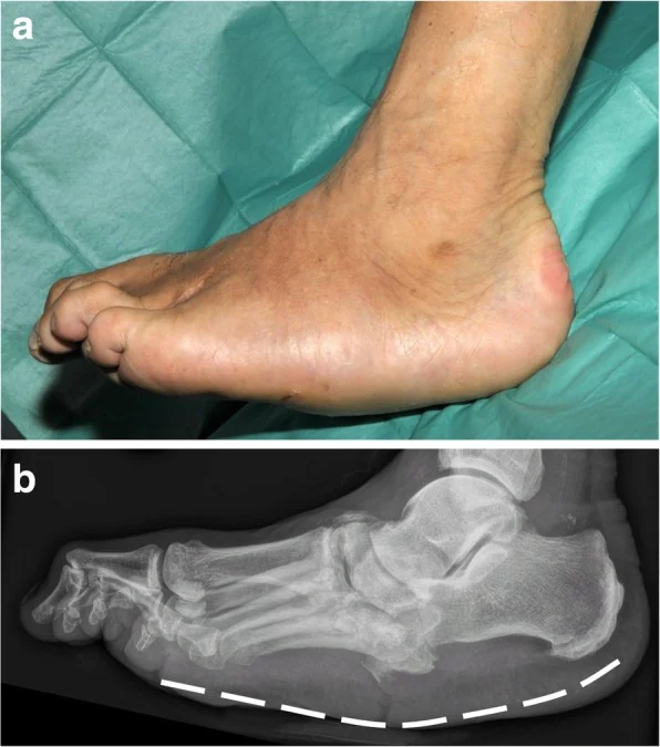
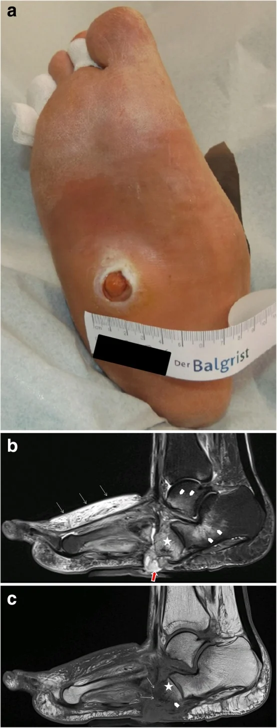
Why it happens
The mechanism is debated but involves:
- Severe neuropathy: the patient can’t feel injury
- Continued walking on the foot: small fractures occur but go unnoticed
- Increased blood flow: neuropathy affects sympathetic regulation, leading to more bone resorption
- Inflammation: a vicious cycle of swelling, weakening, and microfractures
- The bones literally dissolve and fragment under continued weight-bearing
Diagnosis
A red hot swollen foot in someone with diabetes should be assumed Charcot until proven otherwise, because the consequences of missing it are severe.
The workup typically includes:
- Examination: temperature comparison between feet, looking for distinct ulcer/wound source
- X-rays: may show bone changes (often subtle early; can be normal in early Charcot)
- MRI: much more sensitive; differentiates Charcot from osteomyelitis (infection)
- Bone scan in some cases
When x-ray changes do appear, they follow a recognizable pattern by anatomical zone and worsen over weeks to months:

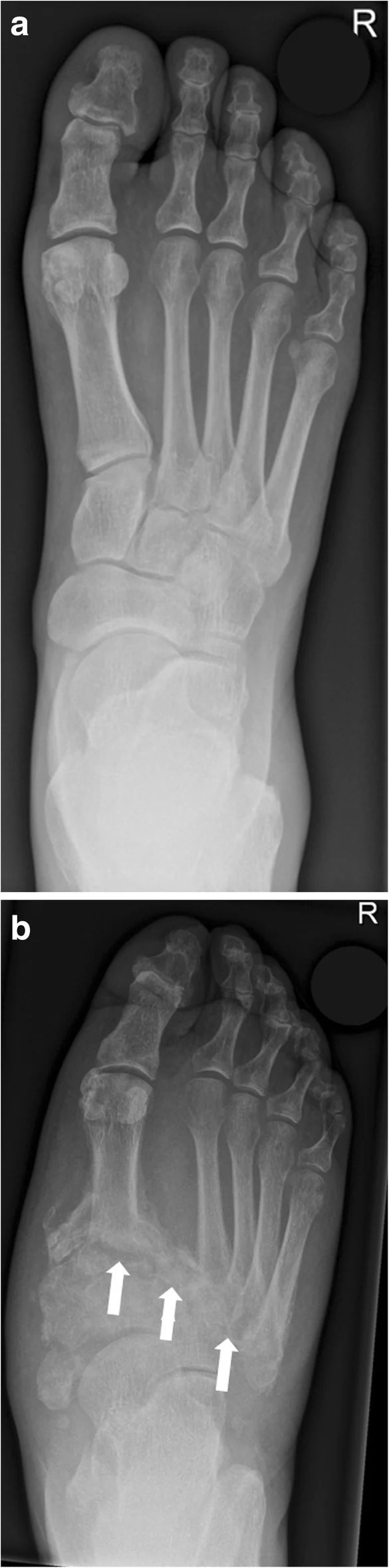
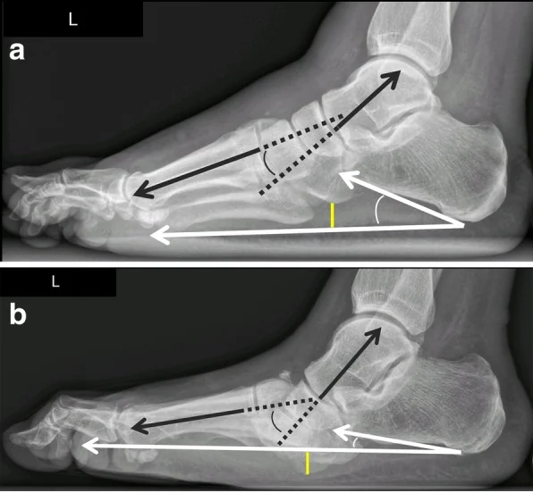
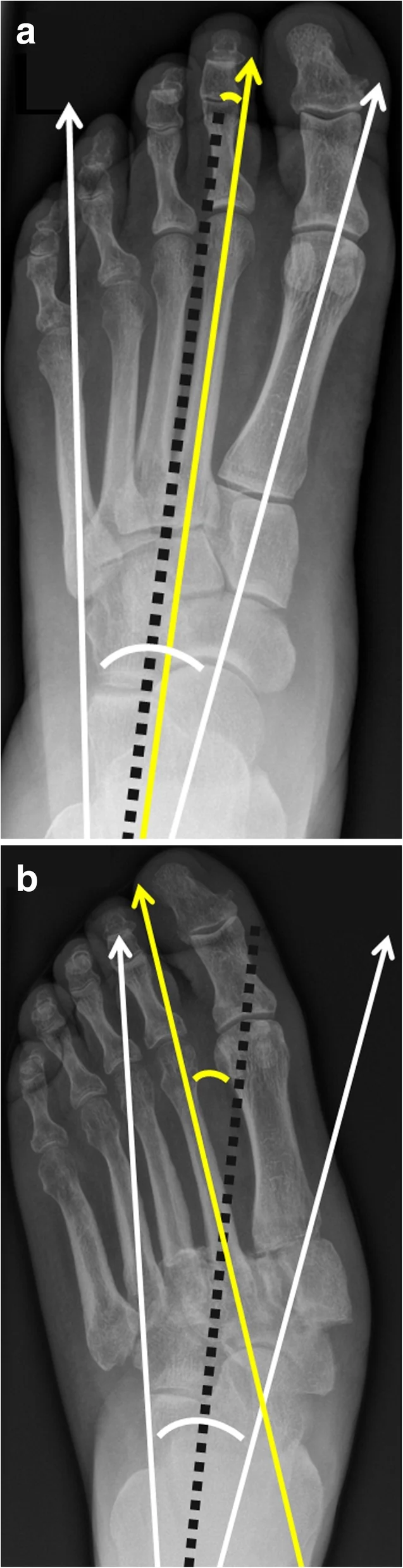
The hardest call: Charcot vs. osteomyelitis (deep bone infection). Both can cause heat, swelling, and bone changes. MRI patterns help; sometimes a biopsy is needed.
MRI can flag Charcot before the x-ray changes, which is why it matters so much for early diagnosis:
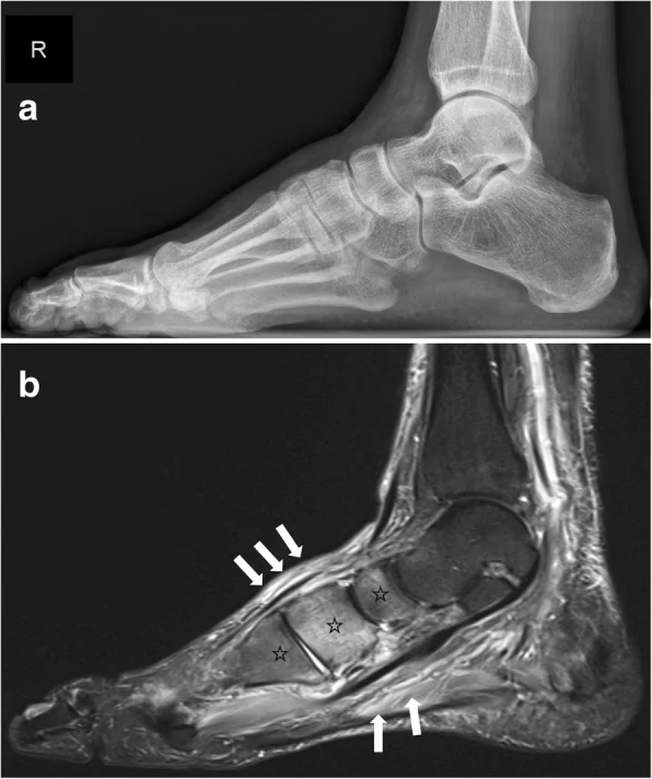
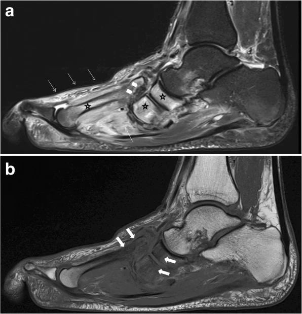
Treatment
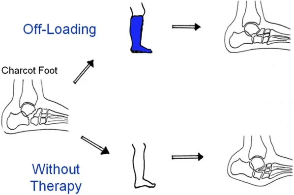
The core principle: immobilize and offload until the inflammation resolves. This often takes 3–6 months or longer.
Active phase
- Total contact cast: the gold standard. Changed weekly. Worn until the foot temperature normalizes and swelling resolves.
- Removable boot (CROW boot): alternative for some patients, but compliance is critical
- Strict non-weight-bearing or limited weight-bearing
- Treatment can take 3–9 months of casting
Coalescent / remodeling phase
- Transition out of the cast into custom shoes or braces
- Ongoing protection and monitoring
Definitive footwear
- Custom-molded diabetic shoes
- CROW boot (Charcot Restraint Orthotic Walker) for severe deformities
- Lifetime protective footwear: Charcot foot remains higher-risk forever
Surgery
- Reserved for failed conservative treatment, severe deformity with skin breakdown risk, or unstable foot that can’t be braced
- Procedures include corrective bone fusion (arthrodesis), often with internal hardware
- Significant operation with prolonged recovery
When to see a clinician
Same day if you have diabetes and:
- A red, hot, swollen foot
- New foot pain or swelling without obvious cause
- Foot that feels warmer than the other side
- Visible deformity developing
This is not a “wait and see” situation.
Prevention
The deeper prevention is preventing severe diabetic neuropathy in the first place, which means glucose control. Once neuropathy is established:
- Daily foot inspection: look for swelling, color changes, temperature differences
- Annual or more frequent podiatry exams
- Properly fitted diabetic shoes
- Avoid barefoot walking: even at home
- Address minor injuries promptly: a sprain in a neuropathic foot needs evaluation
- Awareness: both for patients and primary care doctors. The condition is too often diagnosed late because no one recognizes the early presentation.
Bottom line
Charcot foot is one of the most consequential diabetic foot complications. The single biggest factor in outcomes is how quickly it’s recognized and immobilized. If you have diabetes and develop a red, hot, swollen foot, get it evaluated the same day, even if you don’t think it’s that serious. Especially if you don’t think it’s that serious.
Sources
- Collins AC, Bergman R. Charcot Neuropathic Osteoarthropathy. StatPearls (updated 2026) ↗
- Rogers LC, Frykberg RG, Armstrong DG, et al. The Charcot foot in diabetes. Diabetes Care (2011), ADA/APMA international task force ↗
- American Academy of Orthopaedic Surgeons (OrthoInfo), Diabetic (Charcot) Foot ↗
- Wukich DK, Schaper NC, et al. IWGDF 2023 guidelines on active Charcot neuro-osteoarthropathy in diabetes. Diabetes Metab Res Rev (2024) ↗
- Rosskopf AB, Loupatatzis C, Pfirrmann CWA, Böni T, Berli MC. The Charcot foot: a pictorial review. Insights Imaging (2019); CC BY 4.0 ↗
Last updated: July 16, 2026

About the author
Written and reviewed by a Doctor of Podiatric Medicine (DPM) practicing in Arizona for 6+ years. Board-certified by the American Board of Podiatric Medicine (ABPM); graduate of Midwestern University Arizona College of Podiatric Medicine.
Last clinically reviewed: July 16, 2026