Quick answer
Subtalar arthritis is wear of the cartilage in the joint between the talus and calcaneus — the joint that lets your foot tilt side to side. It’s the most common type of foot arthritis after big-toe arthritis, and the most common cause is prior trauma, particularly a heel (calcaneal fracture) that extended into the joint surface. The classic presentation: deep pain at the back of the foot that hurts most on uneven ground, with stiffness on side-to-side motion.
Why subtalar arthritis develops
The subtalar joint is a complex three-faceted joint that absorbs side-to-side ground forces during walking. Cartilage damage develops in a few main ways:
- Post-traumatic (most common): Calcaneal fractures with intra-articular extension damage cartilage at the time of injury; the damaged surface wears unevenly over years. Talar fractures and severe sprains also drive this pattern.
- Inflammatory arthritis: Rheumatoid arthritis, psoriatic arthritis, and gout can target the subtalar joint along with other small joints.
- Chronic instability: Repeated ankle sprains transfer abnormal forces to the subtalar joint, accelerating cartilage wear.
- Primary osteoarthritis: Less common; happens with age and high cumulative load.
- Following tarsal coalition: When a coalition is partially flexible or after coalition resection, the joint can degenerate over time.
How to recognize it
- Deep pain at the back of the foot, often described as inside the foot rather than at the surface
- Worse on uneven ground — gravel, grass, slopes — because these surfaces demand subtalar motion
- Stiffness with side-to-side motion of the foot (inversion/eversion)
- Morning stiffness that loosens with activity, then aches again at the end of the day
- Vague swelling in the back of the foot below the ankle
- Pain with prolonged standing or walking, less with sitting
- History of a heel or talus fracture, often years earlier
Subtalar arthritis is often mistaken for ankle pain because both are at the back of the foot. The key distinction: ankle pain hurts most with up-down motion; subtalar pain hurts most with side-to-side motion.
Diagnosis
- History and exam — limited subtalar motion, pain on hindfoot inversion/eversion, often with palpable tenderness on the lateral side of the foot below the ankle
- Weight-bearing X-rays — show joint space narrowing, bone spurs, and any old fracture lines. A Broden’s view is a specialized X-ray angle that shows the posterior facet well
- CT scan — gold standard for assessing the joint surfaces, especially when surgery is being considered
- MRI — useful when soft tissue contributions or early disease are suspected
- Diagnostic injection — a small amount of local anesthetic placed into the subtalar joint can confirm that pain truly originates from this joint rather than the ankle or surrounding structures
Treatment
Conservative care (first-line)
Most patients are managed without surgery for years:
- Activity modification — avoid uneven ground when symptomatic; rotate to lower-impact exercise (cycling, swimming)
- NSAIDs for inflammation flares
- Custom orthotics with motion control to reduce subtalar stress
- Stiff-soled shoes or rocker-bottom shoes to limit hindfoot motion
- Ankle-foot orthosis (AFO) or Arizona brace for more advanced cases — limits subtalar motion mechanically
- Physical therapy for balance, stability, and surrounding muscle strength
- Cortisone injection into the subtalar joint can give months of relief and is also diagnostic. Repeated injections are typically limited
Surgery
For pain that fails conservative care:
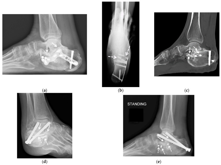
- Subtalar fusion (arthrodesis) — the gold-standard surgery for advanced subtalar arthritis. The joint is permanently fused. Pain relief is usually excellent, but the foot loses side-to-side motion (you can no longer tilt the heel inward or outward).
- Triple arthrodesis — fusion of the subtalar, talonavicular, and calcaneocuboid joints together, used when arthritis affects multiple hindfoot joints
- Recovery — typically 8–12 weeks non-weight-bearing or in a boot, followed by gradual return to walking. Full recovery 6–12 months
- Joint preservation surgery is generally not as durable as fusion for established arthritis
Patients consistently report satisfaction with subtalar fusion when properly indicated — the trade-off of lost motion is usually outweighed by the relief of constant pain.
Bottom line
Subtalar arthritis is overwhelmingly a post-traumatic condition. If you’ve had a calcaneal or talar fracture, expect some degree of subtalar arthritis to develop. Conservative care can keep most patients functional for years; when surgery is needed, subtalar fusion is reliable and well-tolerated. The diagnosis is often delayed because the pain is mistaken for an ankle problem — getting it pinned down early opens up more options.
Last updated: April 27, 2026

About the author
Written and reviewed by a Doctor of Podiatric Medicine (DPM) practicing in Arizona for 6+ years. Board-certified by the American Board of Podiatric Medicine (ABPM); graduate of Midwestern University Arizona College of Podiatric Medicine.
Last clinically reviewed: April 27, 2026