Quick answer
A puncture wound is a deep, narrow injury created by a pointed object piercing the skin. The most common foot scenario: stepping on a nail, splinter, glass, or other sharp object — often through a shoe. The danger isn’t the surface wound, which often looks small and harmless. It’s the depth and contamination: bacteria can be carried deep into tissue, and the small opening doesn’t drain well. Untreated punctures can lead to cellulitis, deep abscesses, and rarely osteomyelitis (bone infection).
What makes puncture wounds different from cuts
A laceration (cut) bleeds out, which helps wash bacteria from the wound. A puncture:
- Doesn’t bleed much — the surface seals quickly
- Doesn’t drain well — bacteria are trapped deep
- May carry foreign material inward — pieces of sock, dirt, rust, the object itself
- Has a deceptively small surface appearance
- Often involves deep structures — tendons, joints, bones — that aren’t visible from outside
This combination is why infections after puncture wounds are common, and why deep infections like osteomyelitis are a real risk.
When to seek care urgently
Most puncture wounds deserve at least a phone call to a clinician. Definitely seek prompt evaluation if:
- The puncture went through a shoe sole — high risk of Pseudomonas infection
- The object is dirty, rusty, or contaminated
- Part of the object may have broken off in the foot
- Tetanus immunization is not up to date (or unknown)
- You have diabetes, peripheral vascular disease, or are immunocompromised
- The wound is deep
- The puncture is over a joint, tendon, or near bone
- You can’t fully clean the wound at home
- There are signs of infection (developing redness, warmth, swelling, pus, fever, red streaks)
- Pain is escalating after the first 24–48 hours rather than improving
Diabetic patients in particular should treat any puncture as worth a clinical evaluation — what looks minor can rapidly become a deep infection.
Initial first aid (within the first hour)
If a clinical evaluation isn’t immediately accessible:
- Wash hands before handling the wound
- Stop bleeding with clean pressure (bleeding helps clean the wound — don’t rush to stop it)
- Remove any easily visible foreign material — but do NOT dig deep or extract embedded objects
- If the object is still embedded and large, leave it in place and seek emergency care — removing it can cause more damage
- Wash thoroughly with clean running water and soap for several minutes
- Apply antibiotic ointment and a clean bandage
- Elevate the foot to reduce swelling
- Avoid weight bearing if possible until evaluated
Don’t:
- Soak in dirty or stagnant water
- Apply hydrogen peroxide repeatedly (damages healing tissue)
- Pour bleach, alcohol, or iodine deep into the wound
- Suture the wound closed at home (sealing in bacteria)
- Ignore the wound — even if it looks minor
Clinical evaluation
A clinician will typically:
- Inspect the wound for depth and contamination
- Image the foot — X-ray is standard for nails, glass, metal; ultrasound or MRI for non-radiopaque objects (wood, plastic, organic material)
- Clean the wound thoroughly — irrigation, sometimes surgical exploration to remove foreign material
- Update tetanus immunization if needed (most adults need a booster every 10 years; sooner for high-risk wounds)
- Consider antibiotics — not all puncture wounds need them, but the threshold is lower for higher-risk wounds
- Arrange follow-up to monitor for infection
Tetanus
Tetanus is a rare but devastating infection caused by Clostridium tetani spores, which are common in soil, dust, and rust. Puncture wounds are classic tetanus-prone injuries because they create the deep, low-oxygen environment the bacteria need.
- If your last tetanus shot was more than 5 years ago for a tetanus-prone wound, get a booster
- If your last tetanus shot was more than 10 years ago for any wound, get a booster
- If you’ve never had tetanus immunization, you need both immune globulin and the vaccine series
When in doubt, get a booster — they’re safe and the consequences of tetanus are severe.
Specific organisms and concerns
Through-the-shoe punctures
A nail through a sneaker is the classic high-risk puncture. The plantar foot environment plus the warm, moist sneaker creates conditions that favor:
- Pseudomonas aeruginosa — the classic organism for through-shoe punctures, especially in the forefoot
- Pseudomonas osteomyelitis — a deep bone infection sometimes following these injuries
Antibiotics chosen for through-shoe punctures often need to cover Pseudomonas specifically.
Diabetic patients
Even minor punctures need close follow-up:
- Higher infection rates
- Slower healing
- Higher risk of osteomyelitis
- Often less pain warning due to neuropathy
- Lower threshold for hospitalization, IV antibiotics, and surgical debridement
Marine and freshwater environments
Punctures sustained in water introduce different organisms:
- Vibrio species in saltwater
- Aeromonas, Mycobacterium marinum in freshwater
- Antibiotic choices may differ from typical soil/shoe punctures
Warning signs of developing infection
Watch the wound for the first 1–2 weeks:
- Increasing redness spreading from the wound
- Warmth to the touch
- Increasing swelling
- Pus at the wound site
- Worsening pain rather than improving
- Red streaks running up the foot toward the ankle
- Fever or chills
- Foul odor
Any of these warrants prompt clinical evaluation.
Real-case example: when a puncture becomes osteomyelitis
A published case report in Cureus (2026) shows exactly why diabetic puncture wounds need aggressive treatment from the start.
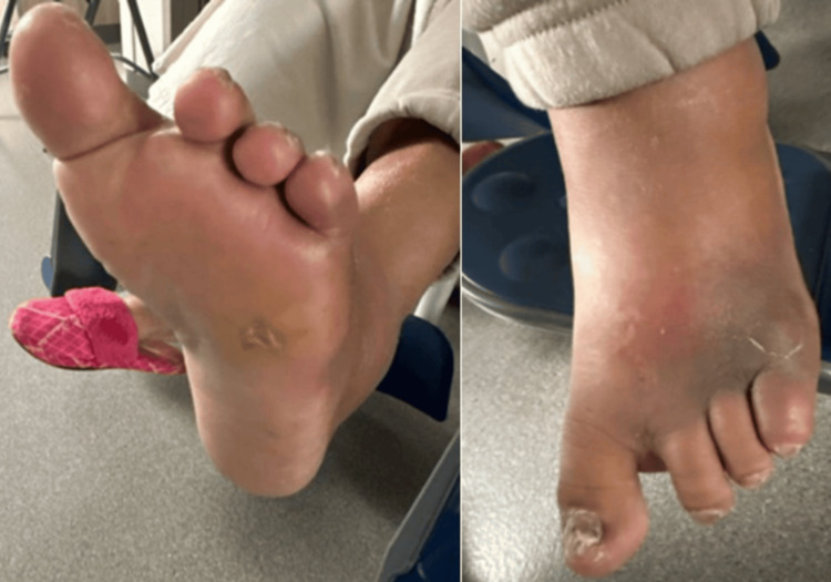
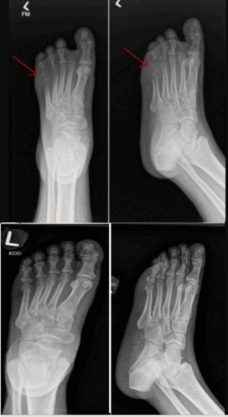
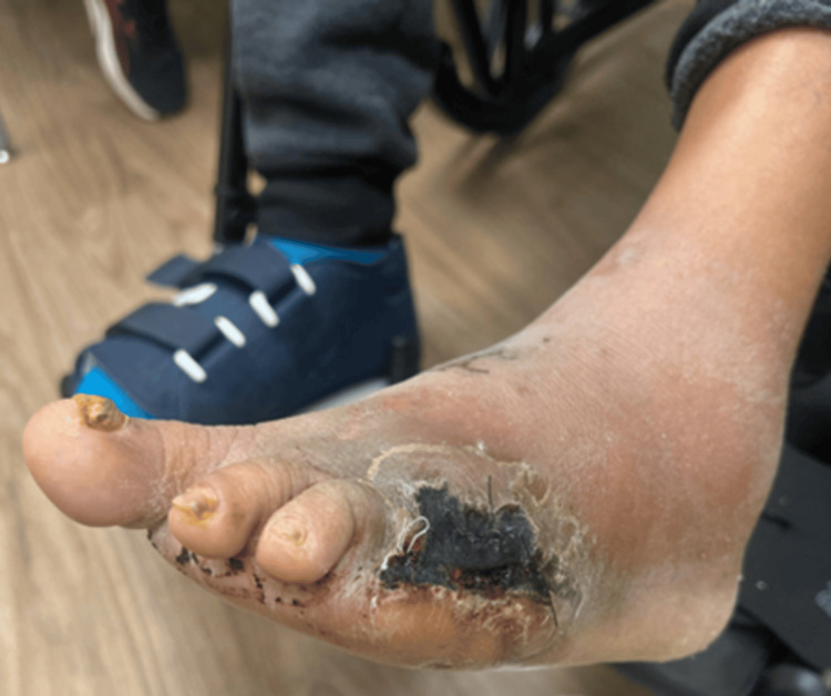
A 62-year-old woman with uncontrolled type 2 diabetes stepped on a nail that went through her shoe and into her forefoot. She was started on outpatient oral antibiotics (cephalexin) for cellulitis, but the infection didn’t respond. Within days, X-rays confirmed MRSA osteomyelitis of the 5th metatarsal — the bone was being destroyed. The course required IV antibiotics and ultimately a ray amputation (surgical removal of the 5th toe along with its metatarsal bone). She recovered, but the foot is permanently changed.
The lesson: in a diabetic patient, a puncture wound that “doesn’t look that bad” can be hiding a deep, rapidly progressing bone infection. Oral antibiotics alone may not stop it. Lower the threshold for X-ray, IV therapy, and surgical evaluation — earlier action prevents amputation.
Click to show clinical photo: the wound at presentation (graphic)
Click to show X-ray: bone destruction (osteomyelitis)
Click to show post-amputation photo (very graphic)
Bottom line
Puncture wounds are deceptive — small on the surface, potentially serious underneath. Wash the wound, ensure tetanus is current, and seek clinical evaluation for any puncture that’s deep, contaminated, through a shoe, in a person with diabetes, or showing any sign of infection. The infections that follow neglected punctures (cellulitis, deep abscess, osteomyelitis) are far more difficult to treat than the original wound — prevention through proper initial care is the right approach.
Sources
Last updated: April 30, 2026

About the author
Written and reviewed by a Doctor of Podiatric Medicine (DPM) practicing in Arizona for 6+ years. Board-certified by the American Board of Podiatric Medicine (ABPM); graduate of Midwestern University Arizona College of Podiatric Medicine.
Last clinically reviewed: April 30, 2026